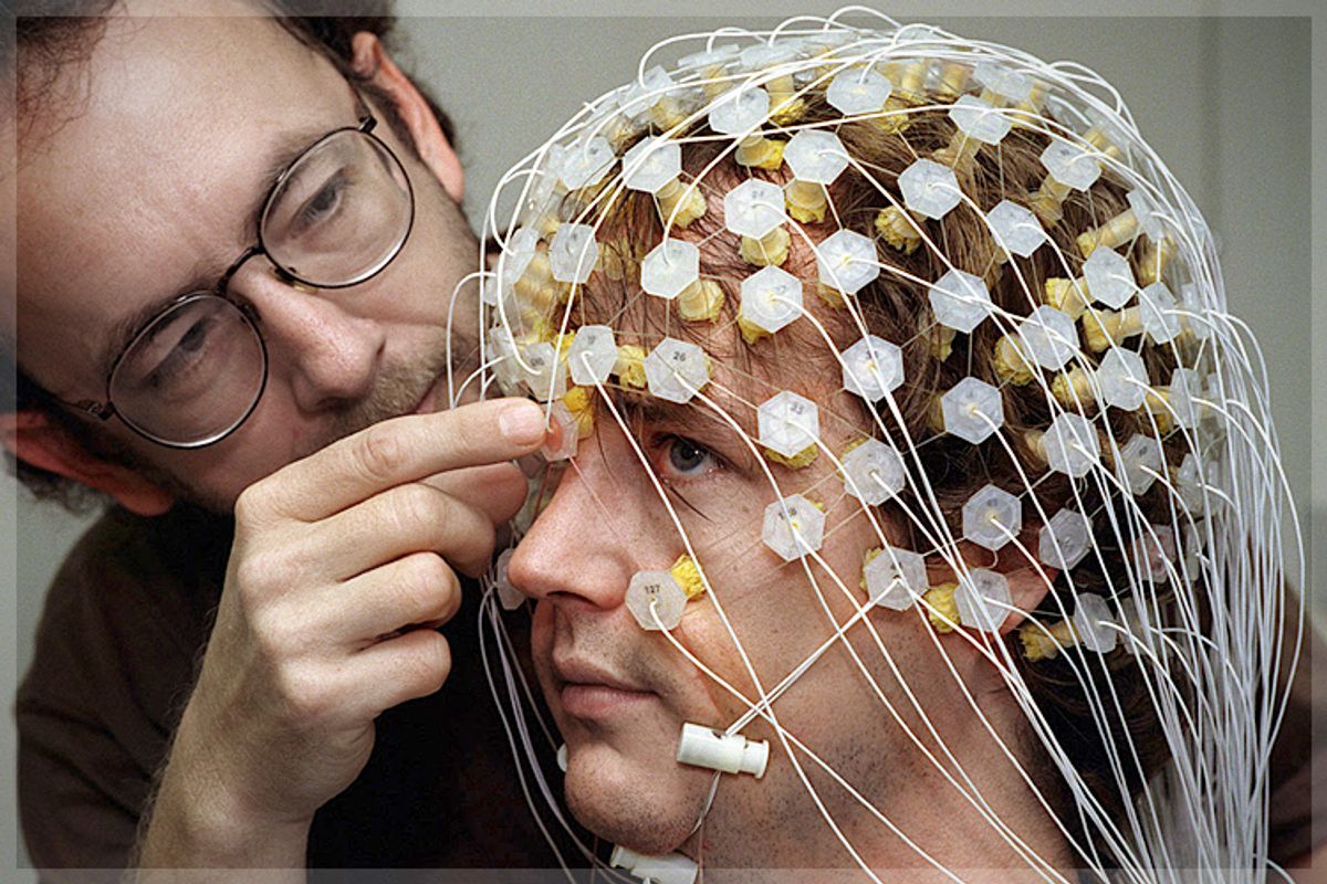
By now you’ve seen the pretty pictures: Color-drenched brain scans capturing Buddhist monks meditating, addicts craving cocaine, and college sophomores choosing Coke over Pepsi. The media—and even some neuroscientists, it seems—love to invoke the neural foundations of human behavior to explain everything from the Bernie Madoff financial fiasco to slavish devotion to our iPhones, the sexual indiscretions of politicians, conservatives’ dismissal of global warming, and even an obsession with self-tanning.
Brains are big on campus, too. Take a map of any major university, and you can trace the march of neuroscience from research labs and medical centers into schools of law and business and departments of economics and philosophy. In recent years, neuroscience has merged with a host of other disciplines, spawning such new areas of study as neurolaw, neuroeconomics, neurophilosophy, neuromarketing, and neurofinance. Add to this the birth of neuroaesthetics, neurohistory, neuroliterature, neuromusicology, neuropolitics, and neurotheology. The brain has even wandered into such unlikely redoubts as English departments, where professors debate whether scanning subjects’ brains as they read passages from Jane Austen novels represents (a) a fertile inquiry into the power of literature or (b) a desperate attempt to inject novelty into a field that has exhausted its romance with psychoanalysis and postmodernism.
Brains are in demand. Once the largely exclusive province of neuroscientists and neurologists, the brain has now entered the popular mainstream. As a newly minted cultural artifact, the brain is portrayed in paintings, sculptures, and tapestries and put on display in museums and galleries.
The prospect of solving the deepest riddle humanity has ever contemplated—itself—by studying the brain has captivated scholars and scientists for centuries. But never before has the brain so vigorously engaged the public imagination. The prime impetus behind this enthusiasm is a form of brain imaging called functional magnetic resonance imaging (fMRI), an instrument that came of age a mere two decades ago, which measures brain activity and converts it into the now-iconic vibrant images one sees in the science pages of the daily newspaper.
As a tool for exploring the biology of the mind, neuroimaging has given brain science a strong cultural presence. As one scientist remarked, brain images are now “replacing Bohr’s planetary atom as the symbol of science.” With its implied promise of decoding the brain, it is easy to see why brain imaging would beguile almost anyone interested in pulling back the curtain on the mental lives of others: politicians hoping to manipulate voter attitudes, marketers tapping the brain to learn what consumers really want to buy, agents of the law seeking an infallible lie detector, addiction researchers trying to gauge the pull of temptations, psychologists and psychiatrists seeking the causes of mental illness, and defense attorneys fighting to prove that their clients lack malign intent or even free will.
The problem is that brain imaging cannot do any of these things—at least not yet.
Why the fixation? First, of course, there is the very subject of the scans: the brain itself. How this enormous neural edifice gives rise to subjective feelings is one of the greatest mysteries of science and philosophy.
Now combine this with the power of human vision. There are good evolutionary reasons for this: The major threats to our ancestors were apprehended visually; so were their sources of food. Plausibly, the survival advantage of vision gave rise to our reflexive bias for believing that the world is as we perceive it to be, an error that psychologists and philosophers call naive realism. This misplaced faith in the trustworthiness of our perceptions is the wellspring of two of history’s most famously misguided theories: that the world is flat and that the sun revolves around the earth. For thousands of years, people trusted their raw impressions of the heavens.
Brain scan images are not what they seem either—or at least not how the media often depict them. They are not photographs of the brain in action in real time. Scientists can’t just look “in” the brain and see what it does. Those beautiful color-dappled images are actually representations of particular areas in the brain that are working the hardest—as measured by increased oxygen consumption—when a subject performs a task such as reading a passage or reacting to stimuli, such as pictures of faces. The powerful computer located within the scanning machine transforms changes in oxygen levels into the familiar candy-colored splotches indicating the brain regions that become especially active during the subject’s performance. Despite well-informed inferences, the greatest challenge of imaging is that it is very difficult for scientists to look at a fiery spot on a brain scan and conclude with certainty what is going on in the mind of the person.
Neuroimaging is a young science, barely out of its infancy, really. In such a fledgling enterprise, the half-life of facts can be especially brief. To regard research findings as settled wisdom is folly, especially when they emanate from a technology whose implications are still poorly understood. As any good scientist knows, there will always be questions to hone, theories to refine, and techniques to perfect. Nonetheless, scientific humility can readily give way to exuberance. When it does, the media often seem to have a ringside seat at the spectacle.
Several years ago, as the 2008 presidential election season was gearing up, a team of neuroscientists from UCLA sought to solve the riddle of the undecided, or swing, voter. They scanned the brains of swing voters as they reacted to photos and video footage of the candidates. The researchers translated the resultant brain activity into the voters’ unspoken attitudes and, together with three political consultants from a Washington, D.C.-based firm called FKF Applied Research, presented their findings in the New York Times in an op-ed titled “This Is Your Brain on Politics.” There, readers could view scans dotted with tangerine and neon yellow hot spots indicating regions that “lit up” when the subjects were exposed to images of Hillary Clinton, Mitt Romney, John Edwards, and other candidates. Revealed in these activity patterns, the authors claimed, were “some voter impressions on which this election may well turn.” Among those impressions was that two candidates had utterly failed to “engage” with swing voters. Who were these unpopular politicians? John McCain and Barack Obama, the two eventual nominees for president.
Another much-circulated study, published in 2008, “The Neural Correlates of Hate” came from neuroscientists at University College London. The researchers asked subjects to bring in photos of people they hated—generally ex-lovers, work rivals, or reviled politicians—as well as people about whom subjects felt neutrally. By comparing their responses—that is, patterns of brain activation elicited by the hated face—with their reaction to the neutral photos, the team claimed to identify the neurological correlates of intense hatred. Not surprisingly, much of the media coverage attracted by the study flew under the headline: “‘Hate Circuit’ Found in Brain.”
One of the researchers, Semir Zeki, told the press that brain scans could one day be used in court—for example, to assess whether a murder suspect felt a strong hatred toward the victim. Not so fast. True, these data do reveal that certain parts of the brain become more active when people look at images of people they hate and presumably feel contempt for them as they do so. The problem is that the illuminated areas on the scan are activated by many other emotions, not just hate. There is no newly discovered collection of brain regions that are wired together in such a way that they comprise the identifiable neural counterpart of hatred.
University press offices, too, are notorious for touting sensational details in their media-friendly releases: Here’s a spot that lights up when subjects think of God (“Religion center found!”), or researchers find a region for love (“Love found in the brain”). Neuroscientists sometimes refer disparagingly to these studies as “blobology,” their tongue-in-cheek label for studies that show which brain areas become activated as subjects experience X or perform task Y. To repeat: It’s all too easy for the nonexpert to lose sight of the fact that fMRI and other brain-imaging techniques do not literally read thoughts or feelings. By obtaining measures of brain oxygen levels, they show which regions of the brain are more active when a person is thinking, feeling, or, say, reading or calculating. But it is a rather daring leap to go from these patterns to drawing confident inferences about how people feel about political candidates or paying taxes, or what they experience in the throes of love.
Pop neuroscience makes an easy target, we know. Yet we invoke it because these studies garner a disproportionate amount of media coverage and shape public perception of what brain imaging can tell us. Skilled science journalists cringe when they read accounts claiming that scans can capture the mind itself in action. Serious science writers take pains to describe quality neuroscience research accurately. Indeed, an eddy of discontent is already forming. “Neuromania,” “neurohubris,” and “neurohype”—“neurobollocks,” if you’re a Brit—are just some of the labels that have been brandished, sometimes by frustrated neuroscientists themselves. But in a world where university press releases elbow one another for media attention, it’s often the study with a buzzy storyline (“Men See Bikini-Clad Women as Objects, Psychologists Say”) that gets picked up and dumbed down.
The problem with such mindless neuroscience is not neuroscience itself. The field is one of the great intellectual achievements of modern science. Its instruments are remarkable. The goal of brain imaging is enormously important and fascinating: to bridge the explanatory gap between the intangible mind and the corporeal brain. But that relationship is extremely complex and incompletely understood. Therefore, it is vulnerable to being oversold by the media, some overzealous scientists, and neuroentrepreneurs who tout facile conclusions that reach far beyond what the current evidence warrants— fits of “premature extrapolation,” as British neuroskeptic Steven Poole calls them. When it comes to brain scans, seeing may be believing, but it isn’t necessarily understanding.
Some of the misapplications of neuroscience are amusing and essentially harmless. Take, for instance, the new trend of neuromanagement books such as Your Brain and Business: The Neuroscience of Great Leaders, which advises nervous CEOs “to be aware that anxiety centers in the brain connect to thinking centers, including the PFC [prefrontal cortex] and ACC [anterior cingulate cortex].” The fad has, perhaps not surprisingly, infiltrated the parenting and education markets, too. Parents and teachers are easy marks for “brain gyms,” “brain-compatible education,” and “brain-based parenting,” not to mention dozens of other unsubstantiated techniques. For the most part, these slick enterprises merely dress up or repackage good advice with neuroscientific findings that add nothing to the overall program. As one cognitive psychologist quipped, “Unable to persuade others about your viewpoint? Take a Neuro-Prefix—influence grows or your money back.”
But reading too much into brain scans matters when real-world concerns hang in the balance. Consider the law. When a person commits a crime, who is at fault: the perpetrator or his or her brain? Of course, this is a false choice. If biology has taught us anything, it is that “my brain” versus “me” is a false distinction. Still, if biological roots can be identified—and better yet, captured on a brain scan as juicy blotches of color—it is too easy for nonprofessionals to assume that the behavior under scrutiny must be “biological” and therefore “hardwired,” involuntary or uncontrollable. Criminal lawyers, not surprisingly, are increasingly drawing on brain images supposedly showing a biological defect that “made” their clients commit murder.
Looking to the future, some neuroscientists envision a dramatic transformation of criminal law. David Eagleman, for one, welcomes a time when “we may someday find that many types of bad behavior have a basic biological explanation [and] eventually think about bad decision making in the same way we think about any physical process, such as diabetes or lung disease.” As this comes to pass, he predicts, “more juries will place defendants on the not-blameworthy side of the line.” But is this the correct conclusion to draw from neuroscientific data? After all, if every behavior is eventually traced to detectable correlates of brain activity, does this mean we can one day write off all troublesome behavior on a don’t-blame-me-blame-my-brain theory of crime? Will no one ever be judged responsible? Thinking through these profoundly important questions turns on how we understand the relationship between the brain and the mind.
Excerpted from "Brainwashed: The Seductive Appeal of Mindless Neuroscience" by Sally Satel and Scott O. Lilienfeld. Published by Basic Books, a member of the Perseus Books Group. Copyright © 2013 by Sally Satel and Scott O. Lilienfeld. Reprinted with permission of the publisher and authors.


Shares