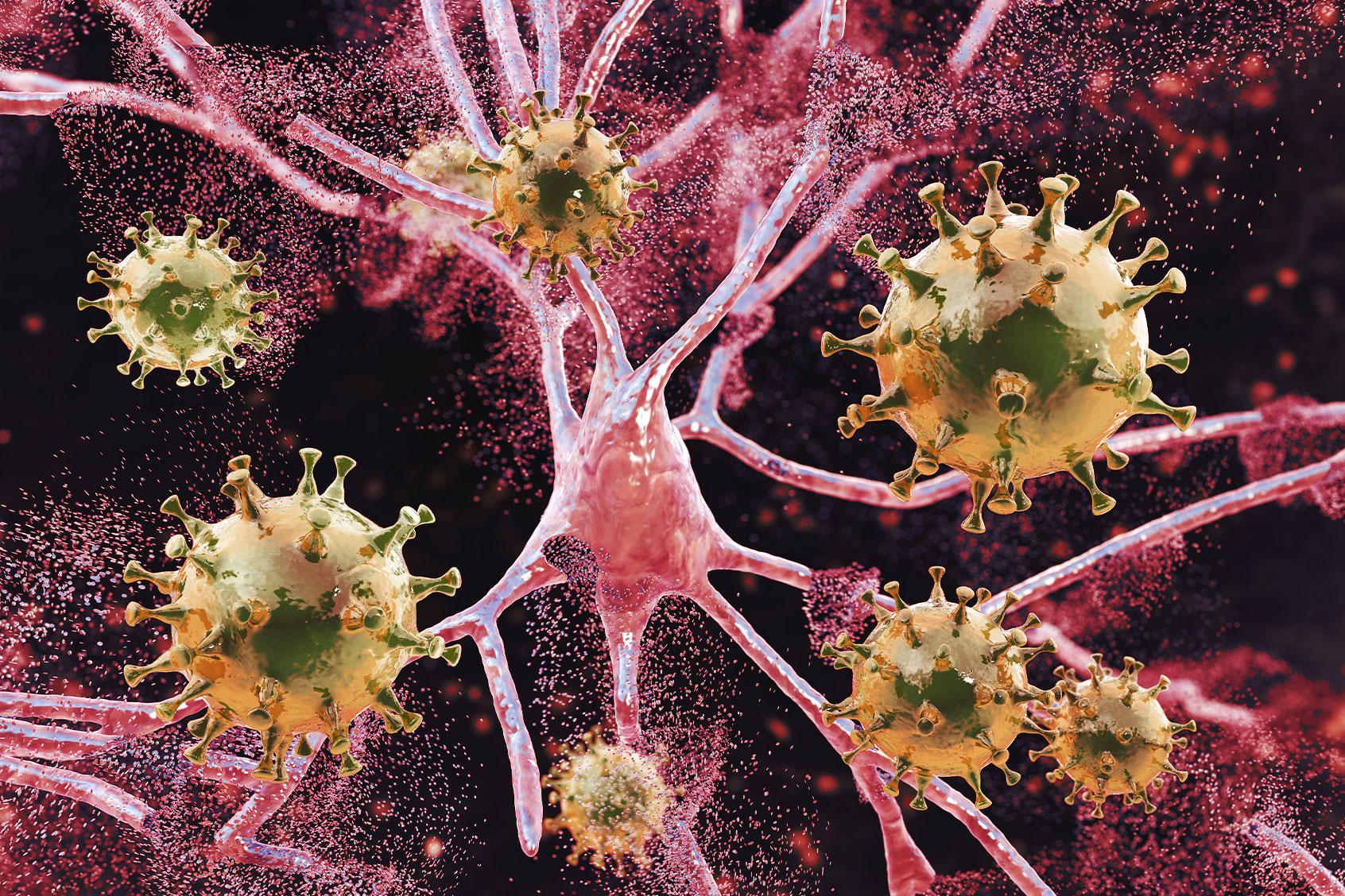
As the pandemic ravaged the globe, it became increasingly clear that COVID, the disease caused by the SARS-CoV-2 virus, does not merely affect the lungs, but it also has effects on the brain. Studies surfaced showing that a COVID-19 infection increased the risk of neurological and psychiatric disorders, caused brain shrinkage and inflammation that mimics Parkinson’s symptoms.
How does that damage manifest in brain scans? Up until now, scientists have only had mediocre images to go by. Thanks to an updated MRI technique, researchers recently got a better look.
“Some may think COVID-19 affects just the lungs,” said Dr. Alexander Wong, a systems design engineering professor at the University of Waterloo. “What was found is that this new MRI technique that we created is very good at identifying changes to the brain due to COVID-19.”
Specifically, through correlated diffusion imaging (CDI), Wong and his colleagues were able to identify that in people who were COVID positive, immediate changes to white matter in two parts of the brain were observed. Previously, Wong invented CDI to detect cancer with better imaging as CDI allows scientists to better see the differences in the ways water molecules move in brain tissue. Since human bodies are made up of a majority of water, understanding how water moves within tissue can provide scientists with information in regards to tissue and its characteristics.
“For example, if water is not flowing, it’s more restricted movement of water, and what that could mean is that the tissue there might see a greater density,” Wong told Salon in an interview.
Want more health and science stories in your inbox? Subscribe to Salon’s weekly newsletter The Vulgar Scientist.
Researchers at Rotman, a world-renowned center for the study of brain function, thought CDI could also be used as a tool to see how COVID could be affecting the brain. In a study published in the journal Human Brain Mapping, Wong and his team looked at the CDI scans of a cohort of people who were either COVID positive or negative. Immediately, they observed differences in the brain scans in the cerebellum and frontal lobe.
COVID-19 can cause changes to the brain — but whether they are damaging or not, is unclear
In the frontal lobe, which is largely responsible for managing thoughts, personality and emotion, there was less restriction of water. In the cerebellum, which is responsible for motor control, there was more restricted diffusion of water molecules.
“In this particular case, when we see more restriction by fusion, that means water is not flowing as well in those particular areas, and essentially, it’s a change,” Wong said. “And so again, what this leads to, we’re not completely sure, but we just observed that the tissue has experienced this change.”
Wong said that the key finding is: “Not only does it affect different areas of the brain, it might affect them differently.”
As to why changes in white matter can be observed in the cerebellum, researchers can only speculate. In the study, Wong and his colleagues suggest that this region of the brain “consistently displays neuropathological abnormalities including immune cell infiltration.” It could be that the cerebellum is very vulnerable to viral entry.
Wong said he hopes one big takeaway from this study is that. as previous research has suggested, COVID-19 can cause changes to the brain — but whether they are damaging or not, is unclear, at least from his study.
“Hopefully, this research can lead to better diagnoses and treatments for COVID-19 patients,” Wong said. “And that could just be the beginning for CDI as it might be used to understand degenerative processes in other diseases such as Alzheimer’s or to detect breast or prostate cancers.”
Read more
about COVID-19


