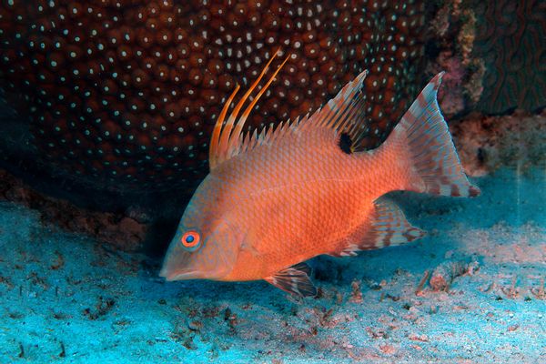
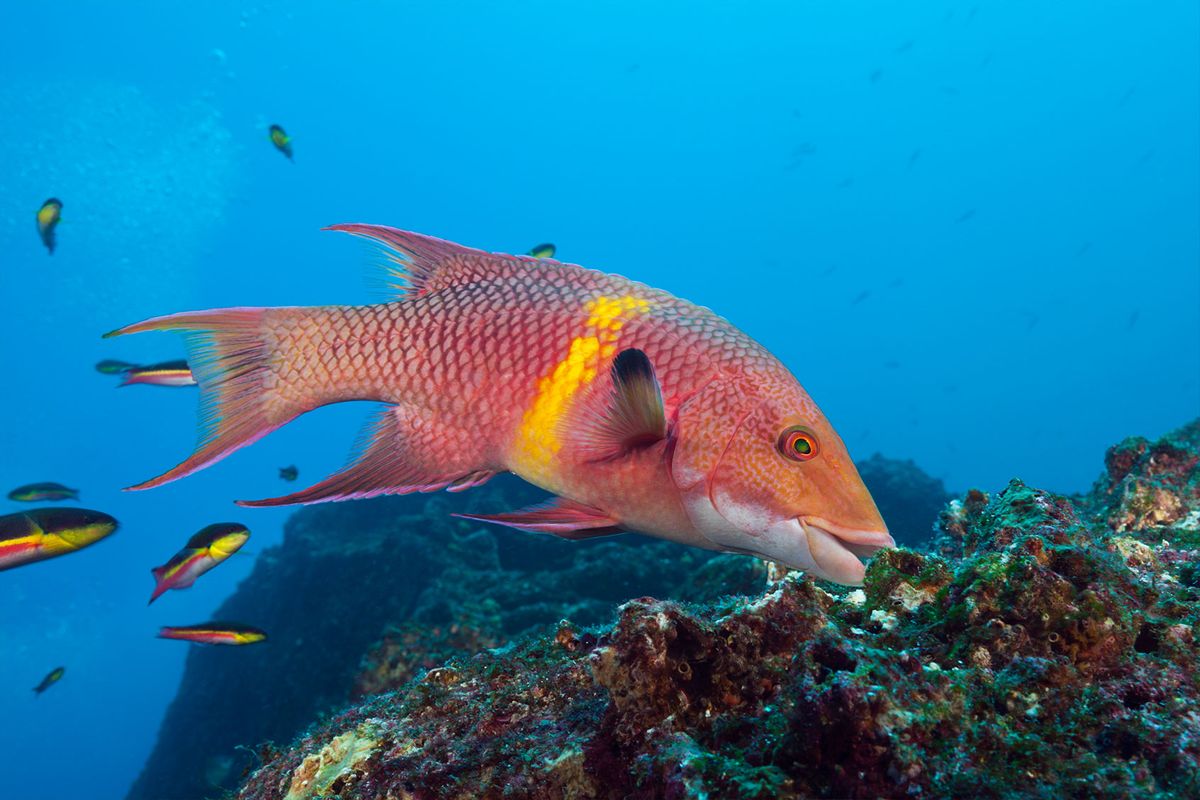
When Lorian E. Schweikert, Ph.D., reeled in a hogfish on a fishing trip to the Florida Keys, she noticed something strange after setting it down on the deck of the boat. Hogfish are known for their camouflaging capabilities, but the one Schweikert captured had transformed its color and texture to match the boat deck — after it was already dead.
"It seemed to be doing it by sensing the environment directly," Schweikert told Salon in a phone interview. "[I thought], 'Maybe the skin can see its environment.'"
Hogfish, octopuses and geckos are some of many color-changing animals that carry light-sensitive proteins called opsins that help them change colors in different environments. It was generally thought opsins functioned as a response to the external environment, but new research conducted by Schweikert — inspired by what she saw on the fishing trip — introduces a new hypothesis: The fish might actually be using the proteins as a sort of internal mirror to monitor themselves.
"It looks like it's watching its own color change performance," Schweikert said.
A hogfish changes color when pigment-containing cells called chromatophores interact with light. When light hits them, they expand or contract, concentrating more or less pigment to create colors. The fish can turn a sandy beige to match the ocean floor and hide or change into flashy colors to scare off predators.
In a 2018 study, Schweikert's team found hogfish possess an opsin specifically sensitive to blue light in their skin, she said. In a study published Tuesday in Nature Communications, they found these blue light receptors were actually buried below the color-changing cells in the fish.
 Seen through a microscope, a hogfish's skin looks like a pointillist painting. (Lori Schweikert, University of North Carolina Wilmington)
"That was surprising because If you can detect blue light with the skin, we thought it would be superficial in the skin, right at the top," Schweikert said. "Instead, we found it highly localized right beneath those color-changing cells."
In other animals with color-changing abilities, color-changing cells or chromatophores are located in the same cell as the animal's light sensors, said Thomas Cronin, Ph.D., a biology professor at the University of Maryland, Baltimore County, who was not involved in this research. Notably, this study showed light sensors were located in different cells than the chromatophores, Cronin told Salon in a phone interview.
 Young hogfish displaying red-mottled phase, Lachnolaimus maximus. (Wild Horizons/Universal Images Group via Getty Images)
"It means, first of all, there has to be some method of communicating through the expansion system how much light is being sensed," Cronin said. "Second, it means in some way, the message from the light absorption system has to go someplace outside of itself — either by hormones or nerves — to get the message back to the filter that it should be expanding or contracting more."
"How would you know you changed color correctly? For these animals, they need a system to know because otherwise, they'd die."
How exactly that communication works isn't clear, but Schweikert hypothesizes that the system is working as a feedback system to keep the camouflage in check. She compared it to our blood pressure changing when we stand up quickly to ensure we don't pass out. The body can sense pressure changes by monitoring or "watching" itself and adjusting appropriately to what it "sees."
"Imagine you had to get dressed in the morning, but you didn't have a mirror and you couldn't bend your neck," Schweikert said. "How would you know you changed color correctly? For these animals, they need a system to know because otherwise, they'd die. They're using color change for camouflage, for thermal regulation, for all of these things. They need to get the color right."
 A pointy-snouted reef fish called the hogfish can change from white to spotted brown to reddish depending on its surroundings. (Photos courtesy of Dean Kimberly and Lori Schweikert)
The process working inside the hogfish is similar to the earliest method of color photography, the autochrome process, in which light travels through plates covered in millions of tiny red, green and blue-colored potato starch grains to produce color.
"The animals can literally take a photo of their own skin from the inside," Duke biologist Sönke Johnsen, one of the study co-authors, said in a statement. However, it's still not clear whether fish can actually "see" this change and there's no evidence that this system is connected to the central nervous system.
"The critical thing the eye has is a lens, which focuses light to create an image," Schweikert said. "But in the skin, without any optical elements that would allow image formation, it doesn't appear dermal photo recognition, or light detected by the skin, is capable of creating images of what's around these animals that they would need to know about [like] what the sea floor looks like or an approaching predator or mate."
We need your help to stay independent
As we increasingly move toward incorporating artificial intelligence into our daily lives, lessons can be drawn about natural processes like these, Schweikert said. Photochromic sunglasses, or glasses that change to sunglasses in sunlight, already use a similar process, Cronin said.
"Whether it's developing smart robots, self-driving cars or other autonomous systems, we can draw principles from natural systems like this to better our own technology," Schweikert said.


Shares